Long-Bao Shi,
Pei-Fu Tang ![]() ,
Wei Zhang,
Yan-Peng Zhao,
Li-Cheng Zhang,
Hao Zhang
,
Wei Zhang,
Yan-Peng Zhao,
Li-Cheng Zhang,
Hao Zhang
For correspondence:- Pei-Fu Tang Email: peifutang@hotmail.com
Received: 3 September 2016 Accepted: 12 December 2016 Published: 31 January 2017
Citation: Shi L, Tang P, Zhang W, Zhao Y, Zhang L, Zhang H. Green synthesis of CuO nanoparticles using Cassia auriculata leaf extract and in vitro evaluation of their biocompatibility with rheumatoid arthritis macrophages (RAW 264.7). Trop J Pharm Res 2017; 16(1):185-192 doi: 10.4314/tjpr.v16i1.25
© 2017 The authors.
This is an Open Access article that uses a funding model which does not charge readers or their institutions for access and distributed under the terms of the Creative Commons Attribution License (http://creativecommons.org/licenses/by/4.0) and the Budapest Open Access Initiative (http://www.budapestopenaccessinitiative.org/read), which permit unrestricted use, distribution, and reproduction in any medium, provided the original work is properly credited..
Purpose: To undertake green synthesis of copper oxide nanoparticles (CuO NPs) using Cassia auriculata leaf extract and evaluate their biocompatibility with rheumatoid arthritis macrophages (RAW 264.7 cell line).
Methods: CuO NPs were prepared by heating a mixture of 10 mL of 0.01 M CuSO4 solution and 30 mL of C. auriculata extract at 80 °C for 1 h. The synthesized CuO NPs were characterized by x-ray diffraction (XRD), Fourier-transform infrared spectroscopy (FTIR), ultraviolet-visible spectroscopy (UV-Vis), energy-dispersive x-ray spectroscopy (EDS), scanning electron microscopy (SEM), transmission electron microscopy (TEM) and dynamic light scattering (DLS). The cytotoxicity of the NPs against RAW 264.7 cells was studied using the 3-(4,5-dimethylthiazol-2-yl)-2,5-diphenyltetrazolium bromide (MTT) assay.
Results: The gradual change in color of the reaction solution from brownish yellow to dark brown indicated CuO NP formation. TEM images revealed spherical, polydispersed NPs (mean particle size, 23 nm). FTIR results indicated capping of polyphenols on the surface of the NPs. Most RAW 264.7 cells (> 95 %) remained alive following exposure to CuO NPs at concentrations of up to 200 μg/mL, indicating biocompatible with the cells.
Conclusion: An eco-friendly, low-cost, biosynthetic method for CuO NP preparation has been successfully developed using C. auriculata leaf extract. Furthermore, the nanoparticles are biocompatible with RAW 264.7 cell line.
Introduction
Copper oxide nanoparticles (CuO NPs) have attracted significant interest due to their wide variety of applications, including in catalysts, gas sensors, high-Tc superconductors, giant magnet resistance materials, solar energy devices, and in the preparation of organic-inorganic nanocomposites [1-5]. Additionally, CuO is used as an antifungal, antibiotic and antimicrobial agent when introduced into textiles and coatings [6].
Moreover, copper-related materials have demonstrated proficient biocidal properties, such as those found in pesticide formulations [7]. Several methods are available for CuO NP preparation, namely, microwave irradiation [8], and sonochemical [9], electrochemical [10] and chemical reduction [11]. However, chemical synthesis of CuO leads to the adsorption of toxic chemicals on its surface, which may have harmful effects when used in biomedical applications. Hence, an environmentally friendly, cost-effective approach is required for CuO NP synthesis. Recently, biosynthesis of nanomaterials using green reducing agents and plants has been reported [12,13]. For example, nanomaterial biosynthesis with extracts of Albizia lebbeck [14], gum karaya [15], Aloe vera [16], and algae [17] has been demonstrated.
The aim of the present work was to synthesize CuO NPs using dried clove extract of Cassia auriculata leaves and evaluate their in vitro cytotoxicity against RAW 264.7 cell line in order to ascertain if they are biocompatible with the cells.
Methods
Materials
Copper sulfate (CuSO4), 3-(4,5-dimethylthiazol-2-yl)-2,5-diphenyltetrazolium bromide (MTT), dimethyl sulfoxide (DMSO), potassium bromide (KBr), and other solvents were purchased from Sigma Aldrich Chemical Ltd., Shanghai, China.
Preparation of plant extract
Leaves of Cassia auriculata were collected from a plant located near the General Hospital of the People’s Liberation Army (No. 28, Fuxing Road, Haiding District, Beijing 100853, China). Approximately 2 g of dried C. auriculata leaves were added to 30 mL of double-distilled water and heated in a water bath for about 2 h at 90°C. The solution was filtered to obtain a clear solution of C. auriculata extract.
Synthesis of CuO nanoparticles
An amount (10 mL) of 0.01 M CuSO4 solution was added to 30 mL of aqueous C. auriculata extract and mixed well with mechanical shaking. The solution was then heated in a water bath at 80 °C for 1 h. The gradual change of color of the reaction solution from brownish yellow to dark brown indicated the formation of CuO NPs.
In vitro cytotoxicity test
RAW 264.7 cells procured from the American Type Culture Collection were initially cultured in RPMI1640 medium containing 1% (w/v) penicillin–streptomycin and 10% (v/v) fetal bovine serum (FBS) at 37°C in a 5 % CO2 atmosphere. For cytotoxicity evaluation, the cells were initially seeded at 1 × 10⁴ cells per well in 96-well titer plates. Cell growth proceeded for 24 h. After two cycles of washing with phosphate buffer solution (PBS; pH: 7.4), various concentrations of CuO NPs were added to each well and incubated for 24 h. After the incubation period, the cells were washed twice with PBS, followed by the addition of fresh culture medium. About 20 μL of MTT reagent (5 mg/mL in PBS) was then added to each well and further incubated for 4 h at 37 °C. The medium in each well was then discarded and DMSO was added to dissolve the cells. The optical density of each well was measured at 570 nm using a microplate reader.
Characterization
XRD analysis was performed using a Bruker D8 Advance diffractometer with Cu-Kα radiation (λ = 1.54 Å) and a step size of 0.02° over the range from 10–80° at a scan rate of about 4°/min. FTIR analysis of the prepared CuO NPs was carried out with a Jasco system to evaluate surface capping. A purified sample was dried and mixed well with KBr powder for pellet preparation for FTIR measurements. The surface morphology and size of the synthesized CuO NPs were analyzed using a high-resolution transmission electron microscope (JEOL JEM 2100 HR-TEM). The sample for TEM analysis was prepared by placing a diluted CuO nanocolloid on the copper grid surface, which was later dried under a vacuum. Additionally, the surface charge and size distribution of the CuO NPs were examined using a Horiba Scientific SZ-100 Nanoparticle Analyzer. Samples for zeta potential analysis were prepared by diluting the CuO NPs with double-distilled water.
Statistical analysis
The results are presented as mean ± standard deviation (SD). Statistical and analytical comparisons of the data were conducted using Student’s t-test or one-way analysis of variance (ANOVA). P < 0.05 was considered to indicate statistical significance.
Results
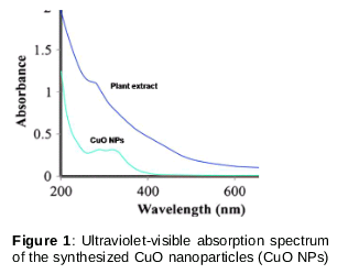
The UV–Vis spectra of aqueous dispersed CuO exhibited a strong surface plasmon resonance (SPR) absorption band at 285 nm, signifying the formation of CuO NPs. No optical absorption peak was observed in the UV-Vis spectra of the plant extract ().
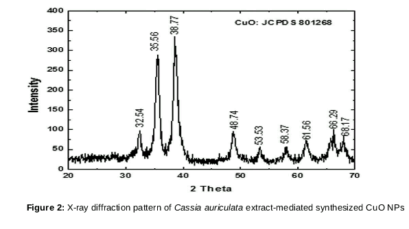
The characteristic XRD peaks observed for CuO NPs prepared using C. auriculata extract are shown in . Intense diffraction peaks at 32.54°, 35.56°, 38.77°, 48.74°, 53.53°, 58.37°, 61.56°, 66.29° and 68.17°, corresponding to 110, 0 02, 111, 202, 020, 202,113, 311 and 113 planes, respectively, indicated the formation of a typical monoclinic CuO NP structure without impurities. Sharp, well-defined CuO reflections in the XRD pattern confirmed the highly crystalline nature of the as-prepared CuO NPs (JCPDS card no. 801268).
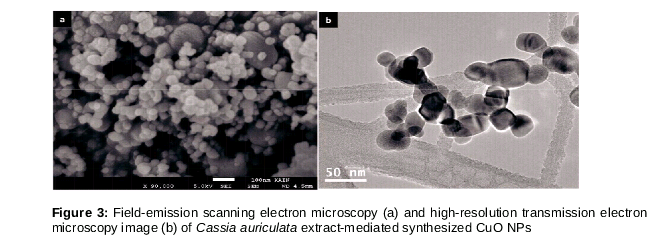
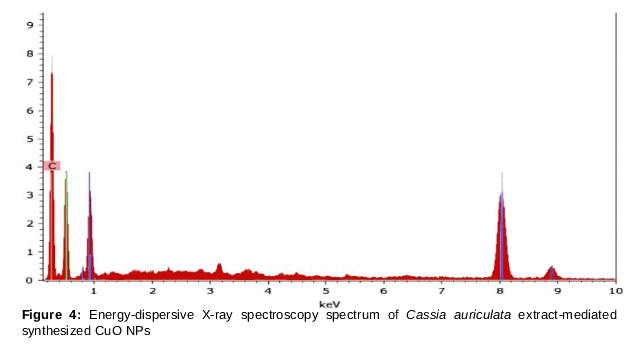
shows field-emission SEM (FE-SEM) and high-resolution TEM (HRTEM) images of the biosynthesized CuO NPs; spherical CuO NP formation was revealed. The average particle size as indicated by TEM was 23.17 nm, in good agreement with the DLS results. The EDS spectrum of the CuO NPs showed the presence of O and Cu peaks only; there was no evidence of impurity peaks (Shown in ).
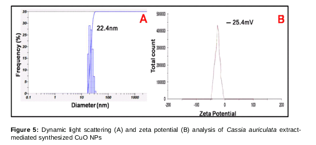
Additionally, DLS analysis was carried out to determine the size distribution of the formed CuO NPs. From A, the average particle size of the CuO NPs was 22.4 nm. Zeta potential analysis showed a negative surface zeta potential of −25.4 mV (B). It is well known that the surface charge and size distribution play key roles in the biological activity of prepared NPs.
shows the presence of bands in the FTIR spectrum of CuO NPs at 3385, 1612, and 1033 cm−1 corresponding to hydroxyl group (-OH) stretching, hydroxyl (-OH) bending, and C-O stretching, respectively. Additionally, the presence of bands at 2922 and 1720 cm−1 is associated with C-H stretching and ketone group vibrations. The bands at 600 and 668 cm−1 are related to Cu-O absorption.
Discussion
The formation of CuO NPs was indicated by the presence of a dark brown precipitate in the bottom of the flask. The Cassia auriculata extract filtrate containing 0.5 M CuSO4 solution began to change color 30 min into the reaction and turned completely dark brown after 1 h. A control experiment performed without the addition of plant extract did not show color change or evidence of CuO NP formation.
The UV-Vis absorption spectrum confirmed CuO NP formation, showing a characteristic SPR band at 285 nm. SPR in the CuO NPs was attributed to the collective oscillation of free surface conduction electrons, excited by the incident electromagnetic radiation. XRD, EDS, TEM analyses indicated the crystalline nature of the plant extract-mediated synthesized CuO NPs. The EDS spectrum confirmed the presence of copper and oxygen; smaller peaks were attributed to biomolecules present in the extract solution. These results are similar to those of previous studies in which green synthesized CuO NPs were prepared using sublimated precursors [18,19].
Furthermore, the zeta potential of the as-synthesized CuO NPs indicated a negative surface charge, which may have been due to the stabilization of the biomolecular constituents of Cassia auriculata extract on the surface of the formed NPs after the reduction process. The negative surface charge of the synthesized CuO NPs further leads to the generation of strong electrostatic repulsion forces among the NPs; this stabilizes the NPs by reducing their aggregation. Notably, the negative surface charge of NPs fabricated using plant extracts has been reported in the literature [20].
The FTIR spectrum of the CuO NPs showed the existence of C-O and hydroxyl functionalities on the NP surface. The FTIR band present at 1720 cm−1 corresponded to ketone functional groups of oxidized polyphenols in the extract of Cassia auriculata, signifying that the synthesized CuO NPs are capped by oxidized polyphenols. Additionally, the stretching bands at 600 and 668 cm−1 are related to Cu-O absorption, further indicating CuO NP formation. These results indicate that the synthesized CuO NPs are stabilized by biomolecular constituents present in the extract of Cassia auriculata.
Macrophages play an important role in rheumatoid arthritis. Synovial fibroblasts along with microphages are involved in the production of a number of matrix-degrading enzymes, chemokines, and cytokines that mediate the interaction with endothelial and inflammatory cells. Subsequently, they are accountable for the successive destruction of bone and articular cartilage, taking into consideration the fact that synovial fibroblasts and macrophages mediate the most significant pathway of joint destruction. Hence, these macrophages would be important targets in current therapeutic approaches to reducing the destruction of bone and cartilage in rheumatoid arthritis [21,22]. In this regard, we have studied the cell viability of RAW 264.7 cell lines after their treatment with CuO NPs to examine the ability of the prepared NPs for drug delivery during the treatment of rheumatoid arthritis.
To study the cytotoxicity of prepared CuO NPs, RAW 264.7 macrophage cell lines were incubated with different concentrations of NPs. Most of the cells (> 95 %) remained alive after exposure to a CuO NP concentration of up to 200 μg/mL, signifying their low cytotoxicity. Thus, CuO NPs prepared using C. auriculata extract may be suitable as a drug delivery vehicle for anti-rheumatic agents for rheumatoid arthritis treatment.
Conclusion
An eco-friendly, low-cost biosynthetic method for the preparation of CuO NPs using Cassia auriculata leaf extract is presented in this work. The Cassia auriculata extract-mediated CuO NPs exhibit very low cytotoxicity towards RAW 264.7 cell lines, thus indicating their biocompatibility with the cells and potential development rheumatoid arthritis therapy.
Declarations
Acknowledgement
References
Archives


News Updates